Neck & Back Pain
Moving beyond imaging to find the true source of spinal pain
What Imaging Misses
MRI reports list findings like "degenerative disc disease" or "mild facet arthropathy," but these same findings appear in people without any pain. The real question isn't what's on the scan. It's what structure is actually generating your pain right now.
Imaging Shows Structure
MRI and X-rays show bones, discs, and joints. They tell you what's there, not what hurts. A bulging disc might be silent. A normal-looking muscle might be the source of everything.
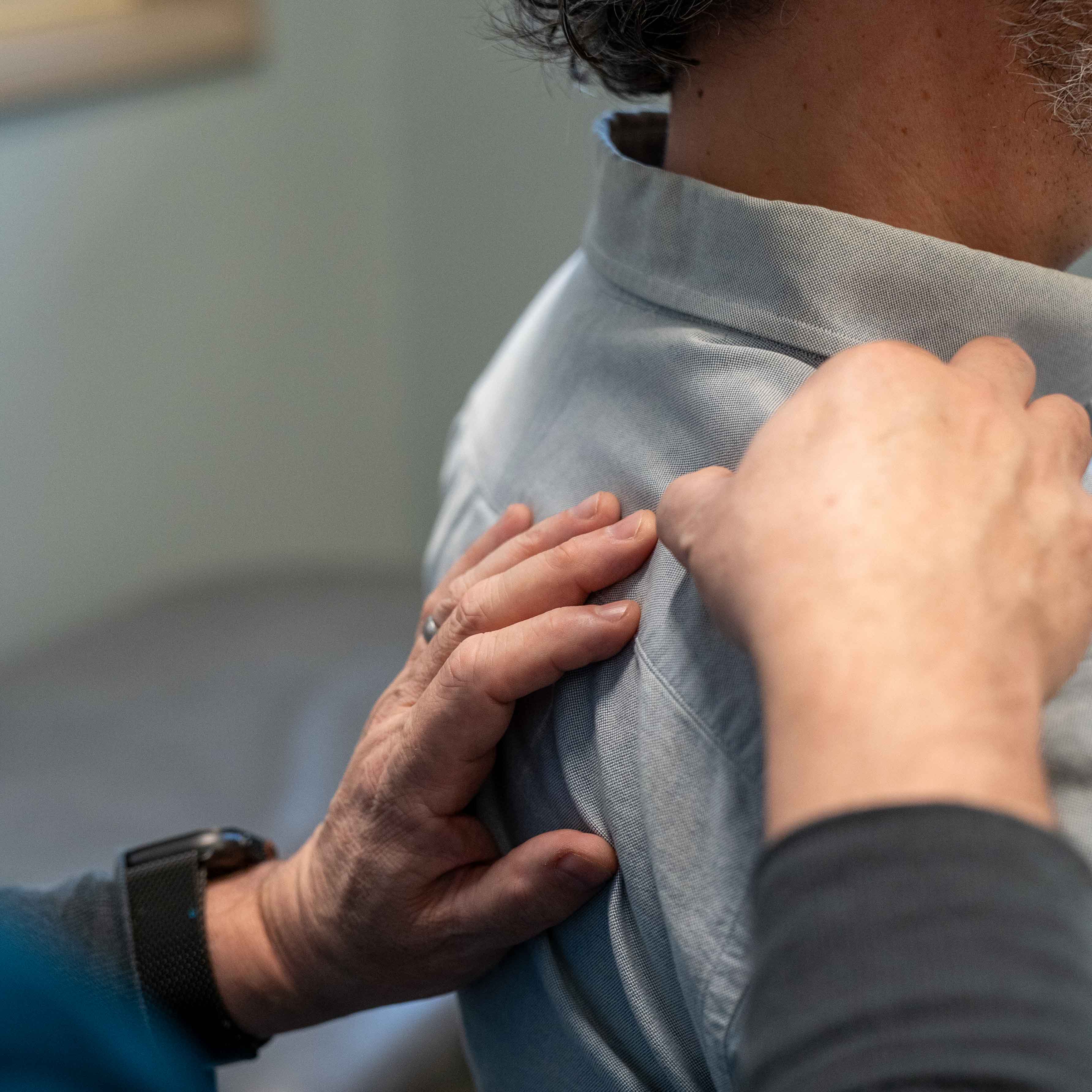
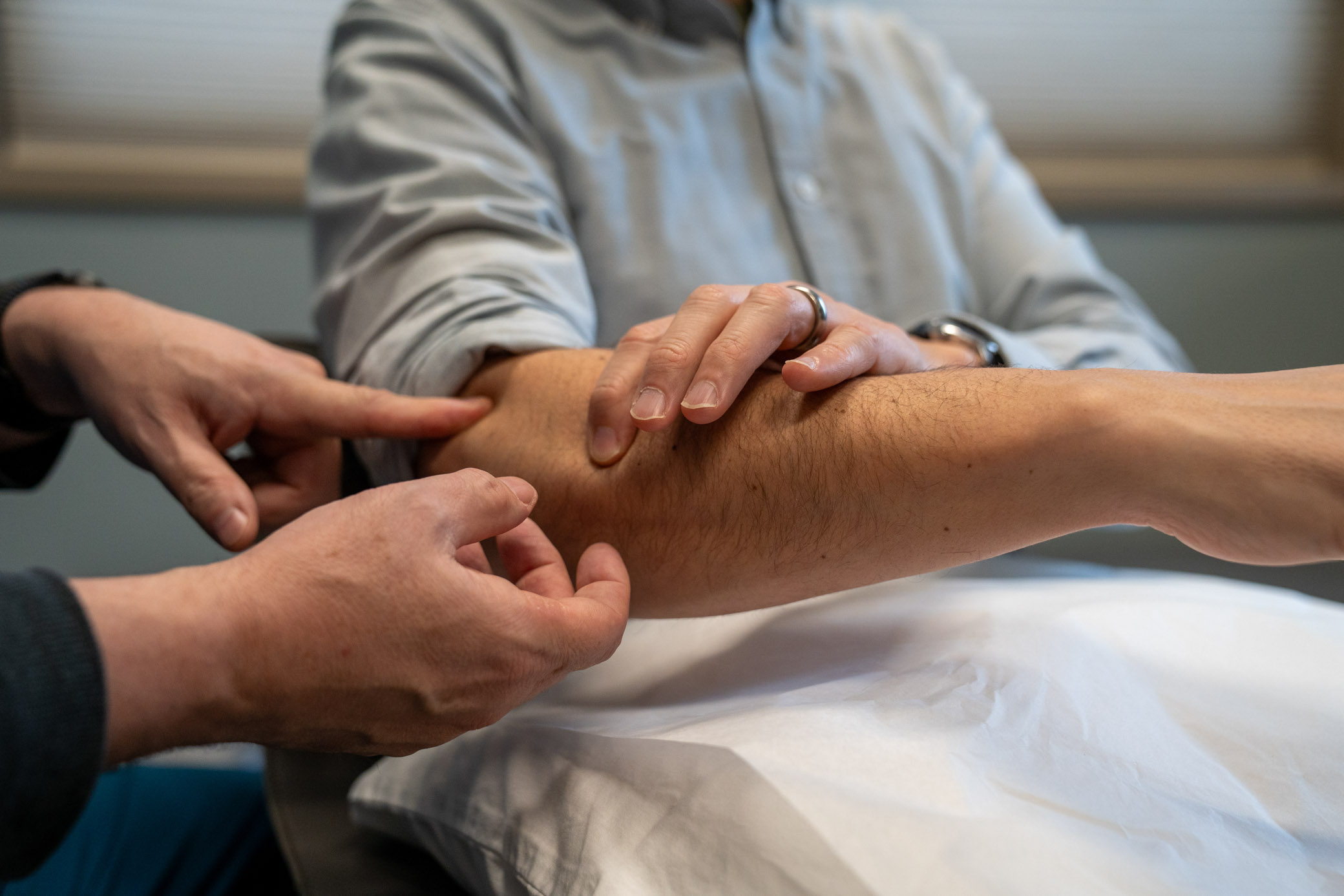
Examination Reveals Function
Pain comes from dysfunction, not just abnormal anatomy. By testing movement, palpating tissues, and correlating your symptoms to specific structures, we find what's actually driving your pain.
Treatment Targets the Source
When we know which structure is generating pain, treatment becomes precise. Calm the irritated facet, release the spastic psoas, or quiet the trigger point, and function returns.
The Unexpectedly Common Sources
After motor vehicle accidents and other trauma, most neck and back pain comes from myofascial trigger points and muscle spasm, not from discs or nerve compression.
Myofascial Referral Patterns
Trigger points in neck and back muscles create referred pain that travels in predictable patterns. Upper trapezius pain radiates into the head. Levator scapulae creates pain at the angle of the neck. Latissimus can send pain down the arm, mimicking nerve compression.
In the lower back, gluteal muscles refer pain down the leg, often mistaken for sciatica. Quadratus lumborum creates deep flank and hip pain. These patterns don't follow dermatomes because they're not coming from nerves.
When we needle these trigger points, the referred pain resolves, often immediately. This is both diagnostic and therapeutic.
The Structural Sources
Cervical Facet Joints
The facet joints in the neck are a common source of post-traumatic pain, though myofascial trigger points causing similar symptoms are even more frequent after motor vehicle accidents. These small joints on the back of each vertebra allow your neck to rotate and tilt. When jarred by whiplash or sudden deceleration, they become inflamed and irritated.
Cervical facet pain typically presents as one-sided neck pain that doesn't cross the midline, often with limited rotation toward the painful side. It can refer pain into the shoulder blade, the base of the skull, or even the front of the shoulder.
Careful examination can identify which level is involved, and medial branch blocks can both diagnose and treat the condition when facet pain is confirmed.
The Psoas & Deep Hip Flexors
The iliopsoas complex attaches directly to the lumbar spine. When this deep hip flexor goes into spasm after injury, it loads the lumbar facet joints and restricts sacroiliac joint mobility, creating persistent lower back pain that doesn't respond to conventional stretching.
Psoas spasm presents as deep lower back pain that worsens with sitting or standing from seated. Patients often describe difficulty finding a comfortable position, and the pain may radiate into the groin or front of the hip.
Treatment focuses on massage and physical therapy to release the deep hip flexor spasm, followed by hip flexor lengthening and glute strengthening to restore balanced mechanics. When the psoas relaxes, facet loading decreases and SI joint mobility improves.
Sacroiliac Joint
The SI joint connects the spine to the pelvis. After motor vehicle accidents, SI joint involvement is common due to seatbelt asymmetry and vertical shear forces during impact. It also occurs after falls, missed steps, or landing heavily on one leg, creating deep buttock pain that radiates down the lateral leg.
SI joint pain is often mistaken for sciatica, but the pain pattern is different. It typically stays more lateral and doesn't travel all the way to the foot. Provocative testing during examination can confirm the diagnosis.
In my experience, a single steroid injection to the SI joint within 1-10 months of injury is often curative when the issue is inflammation. If we determine through examination and injection response that the joint is unstable rather than just inflamed, we transition to prolotherapy or PRP for longer-lasting stabilization.
Lumbar Facet Joints
Like cervical facets, lumbar facet joints can become painful after trauma. However, lumbar facets generate a more diffuse, aching pain in the lower back and buttock, often without the sharp, one-sided character of cervical facet pain.
Lumbar facet pain worsens with extension (arching backward) and twisting. It doesn't radiate below the knee, distinguishing it from true sciatica.
Medial branch blocks can confirm the diagnosis and provide relief, though lumbar facets tend to respond less dramatically than cervical facets.
Disc Herniations (Less Common Than You Think)
Despite what many patients fear, disc herniations are rare after motor vehicle accidents. Biomechanical and epidemiological studies show disc injury occurs in only about 0.01 occupants per 10,000 exposed, typically requiring significant concomitant trauma or unusual loading.
When disc herniations do occur and cause nerve root irritation (radiculopathy), they create dermatomal pain patterns with objective neurologic findings like weakness, reflex loss, or sensory changes. Pain without these findings is almost never from a disc.
Most "bulging discs" on MRI are incidental findings unrelated to current symptoms. The key question is whether the disc is inflaming a nerve root right now, which examination and sometimes EMG can clarify.
How I Find the Source
My evaluation combines detailed movement testing, palpation of specific structures, and correlation with your pain pattern. I'm looking for which structure, when loaded or pressed, reproduces your exact pain.
For myofascial pain, palpating the trigger point recreates the referred pattern. For facet pain, loading the joint in specific planes reproduces the axial pain. For SI joint, provocative maneuvers stress the joint and confirm the diagnosis.
When the clinical picture is clear, imaging confirms anatomy but doesn't change treatment. When it's unclear, imaging helps rule out structural pathology that would require a different approach.
Treatment Matches Source
Once we identify the pain generator, treatment becomes targeted. No shotgun approaches, no hoping something works.
For Myofascial Sources
Trigger point dry needling, graduated stretching, postural correction, and strengthening of opposing muscle groups. Release the spasm, restore balance.
For Facet Joints
Anti-inflammatory approaches, activity modification, and when appropriate, diagnostic/therapeutic medial branch blocks or facet injections. Calm the inflammation, restore movement.
For Psoas Spasm
Massage and physical therapy to release deep hip flexor spasm, followed by hip flexor lengthening and glute strengthening. Restore balanced mechanics to unload the facets and improve SI mobility.
For SI Joint
Steroid injection for inflammatory pain. If examination and injection response reveal joint instability rather than pure inflammation, transition to prolotherapy or PRP for stabilization.
For True Disc Herniation
Time, anti-inflammatory medication, and epidural steroid injections when radicular pain with neurologic findings is present. Most resolve without surgery.
Coordinated Approach
Often, multiple sources contribute. We address them systematically, working with your physical therapist and chiropractor to coordinate timing and intensity of interventions.
Ready to Find What's Actually Wrong?
Let's move beyond the MRI report and figure out which structure is generating your pain. Then we can treat it directly and get you back to normal function.
Schedule Consultation