Joint Pain & Stiffness
Finding and treating the source of restricted movement after trauma
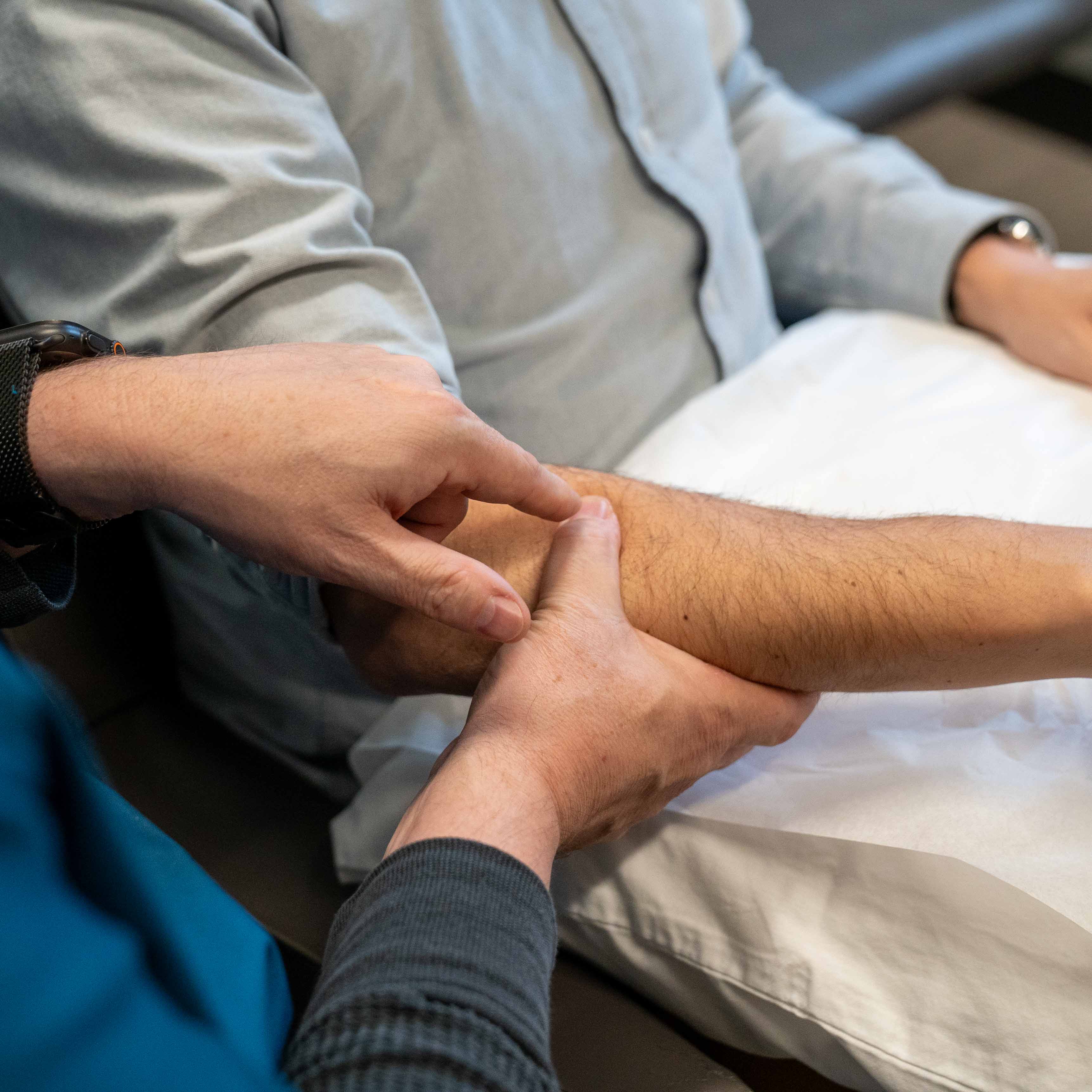
When Joints Stop Moving Normally
After trauma, joints can become stiff, painful, and restricted. The question isn't whether something hurts. It's which structure is inflamed, unstable, or mechanically impaired, and what will restore normal movement.
Spinal Joints
Facet joints and the sacroiliac joint are common pain generators after motor vehicle accidents. These deep structures create localized pain and referred patterns that don't show up on standard imaging but respond well to targeted treatment.
Peripheral Joints
Shoulders, elbows, wrists, hips, knees, and ankles can all be injured during impact or bracing. Inflammation, capsular tightness, and post-traumatic stiffness limit function and create persistent discomfort.
Joint vs. Ligament
Sometimes the issue isn't the joint surface itself but the supporting ligaments. Instability creates different symptoms than inflammation and requires different treatment. Sorting this out guides whether we calm inflammation or stabilize laxity.
Spinal Joints After MVA
Facet and sacroiliac joint pain is very common with motor vehicle accident injuries. These deep structures cause real trouble but respond well to fluoroscopically guided treatment.
Cervical & Lumbar Facets
The small joints on the back of each vertebra allow rotation, flexion, and extension. When jarred by sudden deceleration, they become inflamed and irritated, creating axial neck or back pain that's often one-sided.
Cervical facet pain can refer into the shoulder blade, base of the skull, or even the front of the shoulder. Lumbar facet pain creates a more diffuse ache in the lower back and buttock.
Diagnosis comes from examination, loading the joint in specific planes to reproduce the pain. When conservative care fails, medial branch blocks both diagnose and treat the condition.
The Sacroiliac Joint
The SI joint connects the spine to the pelvis. After motor vehicle accidents, SI joint involvement is common due to seatbelt asymmetry and vertical shear forces during impact.
SI joint pain presents as deep buttock pain that radiates down the lateral leg, often mistaken for sciatica. Unlike true sciatica, it typically stays more lateral and doesn't travel all the way to the foot.
In my experience, a single corticosteroid injection to the SI joint within 1-10 months of injury is often curative when the issue is inflammation. If examination and injection response reveal joint instability, we transition to prolotherapy or PRP for stabilization.
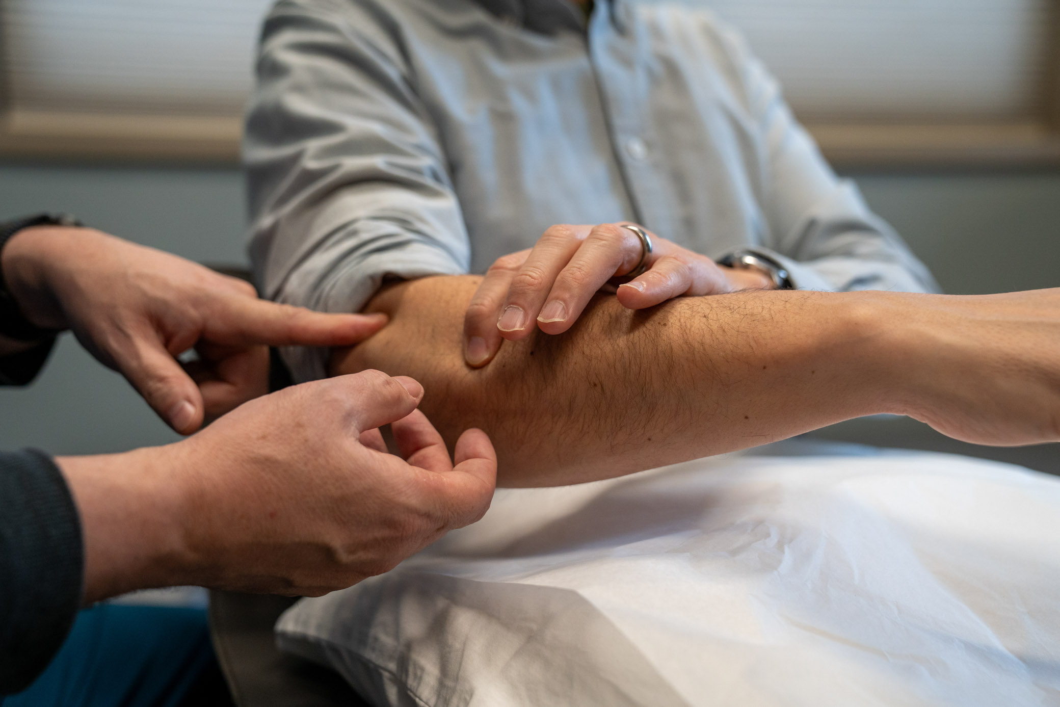
Peripheral Joint Issues
Shoulder Pain & Stiffness
The shoulder is commonly injured during bracing or impact. The glenohumeral joint, acromioclavicular joint, and rotator cuff can all be damaged, creating pain with overhead movement, reaching behind the back, or lifting.
Post-traumatic adhesive capsulitis (frozen shoulder) can develop if early inflammation isn't addressed, leading to progressive stiffness and loss of range of motion.
Treatment focuses on reducing inflammation early, then gradually restoring mobility through targeted physical therapy. Steroid injections can calm acute flares when conservative care stalls.
Elbow, Wrist & Hand Joints
Bracing during collision can strain elbow and wrist joints. Lateral epicondylitis (tennis elbow), medial epicondylitis (golfer's elbow), and wrist ligament sprains create grip weakness and pain with use.
Small joint injuries in the hand and fingers often go unrecognized initially but can lead to chronic stiffness and dysfunction if not properly rehabilitated.
Treatment matches the structure involved. Tendon issues require eccentric loading and progressive strengthening. Joint capsule tightness needs mobilization. Ligament laxity may benefit from prolotherapy.
Hip Joint Pain
Hip joint pain after trauma typically presents as groin pain with walking or hip flexion. This is distinct from trochanteric bursitis (lateral hip pain) or SI joint pain (posterior).
True hip joint pathology may involve labral tears, capsular inflammation, or post-traumatic arthritis. Examination with specific provocative maneuvers and sometimes imaging helps distinguish these.
Conservative management includes activity modification, anti-inflammatory approaches, and physical therapy. Intra-articular hip injections can both diagnose and treat when the source is unclear.
Knee & Ankle Joints
Knee injuries from bracing or dashboard impact can damage meniscus, ligaments, or cartilage. Swelling, instability, and mechanical symptoms (catching, locking) suggest internal derangement.
Ankle sprains are common but can lead to chronic instability if ligaments don't heal properly. Recurrent "giving way" or persistent lateral ankle pain suggests ligamentous laxity rather than simple inflammation.
For instability, prolotherapy or PRP can stimulate ligament healing and restore joint stability. For mechanical issues like meniscal tears, orthopedic referral may be appropriate after conservative trial.
Distinguishing Trauma from Degeneration
X-rays and MRIs often show "degenerative changes" that existed before the accident. The key question: is this causing your current symptoms, or is it an incidental finding?
Degenerative Changes Are Common
By age 40, most people have some degree of joint degeneration on imaging. Mild facet arthropathy, disc degeneration, and osteophytes (bone spurs) are normal age-related changes that often cause no symptoms.
The presence of these findings doesn't mean they're the source of your post-traumatic pain. What matters is whether the joint becomes symptomatic after trauma.
Examination helps distinguish. If loading the joint reproduces your exact pain, it's a pain generator. If pressing nearby muscles recreates the pain, it's myofascial. If neither reproduces it, we keep looking.
Treatment Approaches
Once we identify which joint is the problem, treatment becomes targeted. Inflammation needs calming. Instability needs stabilization. Mechanical restrictions need mobilization.
For Inflammation
Anti-inflammatory approaches, activity modification, physical therapy to maintain range of motion. When conservative care stalls, corticosteroid injections can calm acute inflammation and break the pain cycle.
For Instability
Ligamentous laxity doesn't respond to steroids. Instead, prolotherapy or PRP stimulates tissue healing and stabilizes the joint. This works for SI joint instability, ankle sprains, and other ligament injuries.
For Stiffness
Joint mobilization, progressive stretching, and strengthening opposing muscle groups. The goal is to restore full range of motion and prevent permanent restriction.
Fluoroscopic Guidance
For deep joints like facets and SI joints, I use real-time x-ray guidance to ensure precise needle placement. This isn't guesswork. The medication goes exactly where it needs to be.
Ready to Restore Normal Movement?
Let's identify which joint is the problem, whether it's inflamed or unstable, and treat it appropriately so you can move without restriction again.
Schedule Consultation